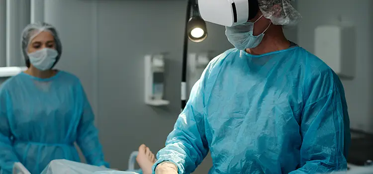
Application of Virtual Reality (VR) Application to Oral Surgery
Cysts and tumors can form in the bones of the oral and jaw, and these diseases are treated by oral surgery. Surgical removal or excision including the jawbone is common, but it is difficult to observe the jawbone directly because it is covered by gums, muscles, and skin.
In addition, disease may also contact or adhere to blood vessels or nerves in or around the jawbone, which can lead to unexpected complications and accidents.
By applying VR (virtual reality) technology, it is possible to reproduce 3D images (Fig. 1) created from the patient’s image data in a VR space (Fig. 2), and the location of cysts and tumors, their size and shape, and their location in relation to surrounding blood vessels and nerves can be observed and understood from various directions in 3D. By sharing such information with medical staff, it will be possible to plan and execute safer and more reliable surgeries.


Augmented Reality (AR) Application to Oral Surgery
In oral surgery that deals with the jawbone, surgical plans are made based on the patient’s image data, and surgical simulations are performed. For example, for patients with jaw deformities, such as a opposite bite or facial asymmetry, we perform surgery to correct facial distortion by cutting the jaw bone and moving it millimeter by millimeter to create normal bite.
Until now, the amount of jaw bone to be moved was calculated based on the patient’s image data, and the surgeon actually measured the amount of movement during the surgery to reproduce it. By applying augmented reality (AR) technology, the surgeon can superimpose the previously simulated osteotomy lines and movement information on the surgical field in three dimensions without taking their eyes off the patient during the surgery.
In addition, blood vessels and nerves that exist inside the bone cannot be observed from outside the bone during surgery, so until now, surgeons have had to perform surgery with image data in their heads to avoid damaging blood vessels and nerves. Using AR, the location of blood vessels and nerves can be superimposed on the surgical field, allowing for safer and more precision surgery.

